 My Account
 +7 (495) 129 37 00
 st. Derbenevskaya, 9
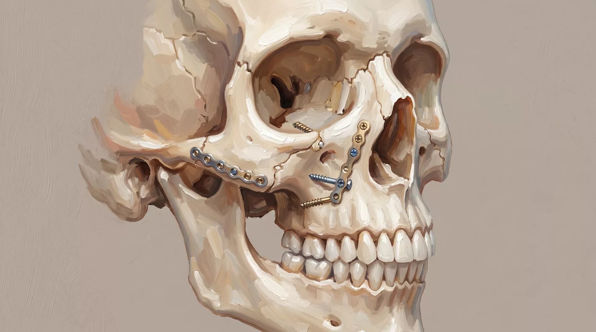
Treatment of Facial Skeleton Fractures (Lower and Upper Jaw, Zygoma, Orbit)
Restoration of proper bite and facial symmetry after fractures using modern titanium plates and 3D tomography.
Available sections:
Treatment of Facial Bone Fractures
Lower Jaw, Upper Jaw, Cheekbones, and Orbit
Fractures of the facial skeleton are not only bone injuries, but also a threat to function: the bite may be disturbed, mouth opening may become difficult, diplopia may occur with orbital fractures, and nasal deformity may develop with midface fractures. Timely surgical treatment in Moscow makes it possible to restore facial anatomy and regain lost functions, provided that the operation is performed technically correctly and within the optimal timeframe.
Types of Fractures and Their Features
Lower Jaw Fractures
The lower jaw is the most commonly fractured bone of the facial skeleton. Typical locations include the condylar process, or articular process, the angle of the jaw, the body, and the symphysis.
Fractures of the condylar process are especially important: if treated incorrectly, they may lead to limited mouth opening and ankylosis, or immobility of the joint. When fragments are displaced, restoration of the bite is possible only surgically.
Upper Jaw Fractures
Upper jaw fractures are classified according to Le Fort types I, II, and III, depending on the fracture level.
Le Fort II and III fractures are accompanied by significant displacement of the midface and require stabilization of several structures at the same time. They are often combined with fractures of the nasal bones and orbit.
Fractures of the Zygomatic Bone and Zygomatic-Orbital Complex
The zygomatic bone connects with four other bones, so its fracture usually involves several articulations.
Clinically, this may appear as depression of the cheekbone area, difficulty opening the mouth when a fragment is displaced toward the coronoid process of the lower jaw, and numbness of the cheek and upper lip.
With timely treatment, anatomy can be fully restored.
Orbital Wall Fractures
A blow-out fracture of the inferior or medial orbital wall occurs after blunt trauma to the eye area.
Orbital fat and the inferior rectus muscle of the eye may become entrapped in the fracture area. This causes diplopia, or double vision, and restricted movement of the eyeball.
Surgery eliminates the entrapment and restores the integrity of the orbital wall.
Diagnostics
The mandatory examination is CT imaging of the facial skeleton with three-dimensional reconstruction. It allows the surgeon to accurately assess the number and position of fragments, the degree of displacement, and the condition of surrounding structures.
Based on the CT findings, the surgeon plans the surgical approaches and the scope of fixation.
Surgical Treatment
The operation is performed under general anesthesia. The goal is repositioning, meaning precise alignment of the fragments in the anatomically correct position, and their reliable fixation.
The surgical approaches are selected according to the fracture location and the need to minimize visible scars: intraoral approaches, through the mucosa, or through natural facial folds or the eyebrow area.
For lower jaw fractures, intermaxillary fixation, or splinting, may be used during the healing period, or open reduction with osteosynthesis may be performed. The choice of method depends on the location and nature of the fracture.
Timing and Rehabilitation
The optimal timing for surgery is within the first 5–10 days after trauma.
After osteosynthesis of the lower jaw, a soft diet is prescribed for 4–6 weeks. Bone healing takes 4–8 weeks depending on the nature of the fracture and the patient’s age.
A follow-up CT scan is performed after 3–4 months.
Contraindications
Contraindications include severe combined injuries in an unstable condition, in which surgery is postponed until stabilization, active infectious processes in the fracture area, and severe systemic diseases in the stage of decompensation.
The final decision is made by the surgeon.
Frequently Asked Questions
Is Surgery Needed for a Jaw Fracture Without Displacement?
For non-displaced fractures, conservative treatment may be possible, including splinting and a soft diet.
If there is displacement, bite disturbance, or instability of the fragments, surgery is indicated. This is determined based on CT findings.
Are the Plates Removed After Osteosynthesis?
In most cases, no. Titanium plates remain permanently.
Removal is indicated in cases of infection, palpable discomfort, or special anatomical situations, as determined by the surgeon.
How Long After Surgery Should I Avoid Solid Food?
For lower jaw fractures, a soft diet is prescribed for 4–6 weeks.
The exact timing depends on the location and complexity of the fracture. The surgeon provides individual recommendations.
Will Sensitivity Recover After a Cheekbone Fracture?
With zygomatic bone fractures, numbness of the cheek and upper lip often occurs due to stretching or compression of the infraorbital nerve.
After repositioning of the fragments, sensitivity usually recovers within several weeks or months.
Book a Consultation
Book a consultation. The surgeon will review the images and recommend a treatment plan.
OUR SPECIALISTS
A team that continues the traditions of Professor A.I. Nerobeev's school. Our specialists not only practice but also teach, developing unique techniques in reconstructive and aesthetic medicine.




Leave your number and we will contact you