 My Account
 +7 (495) 129 37 00
 st. Derbenevskaya, 9
Revascularized Bone Grafting
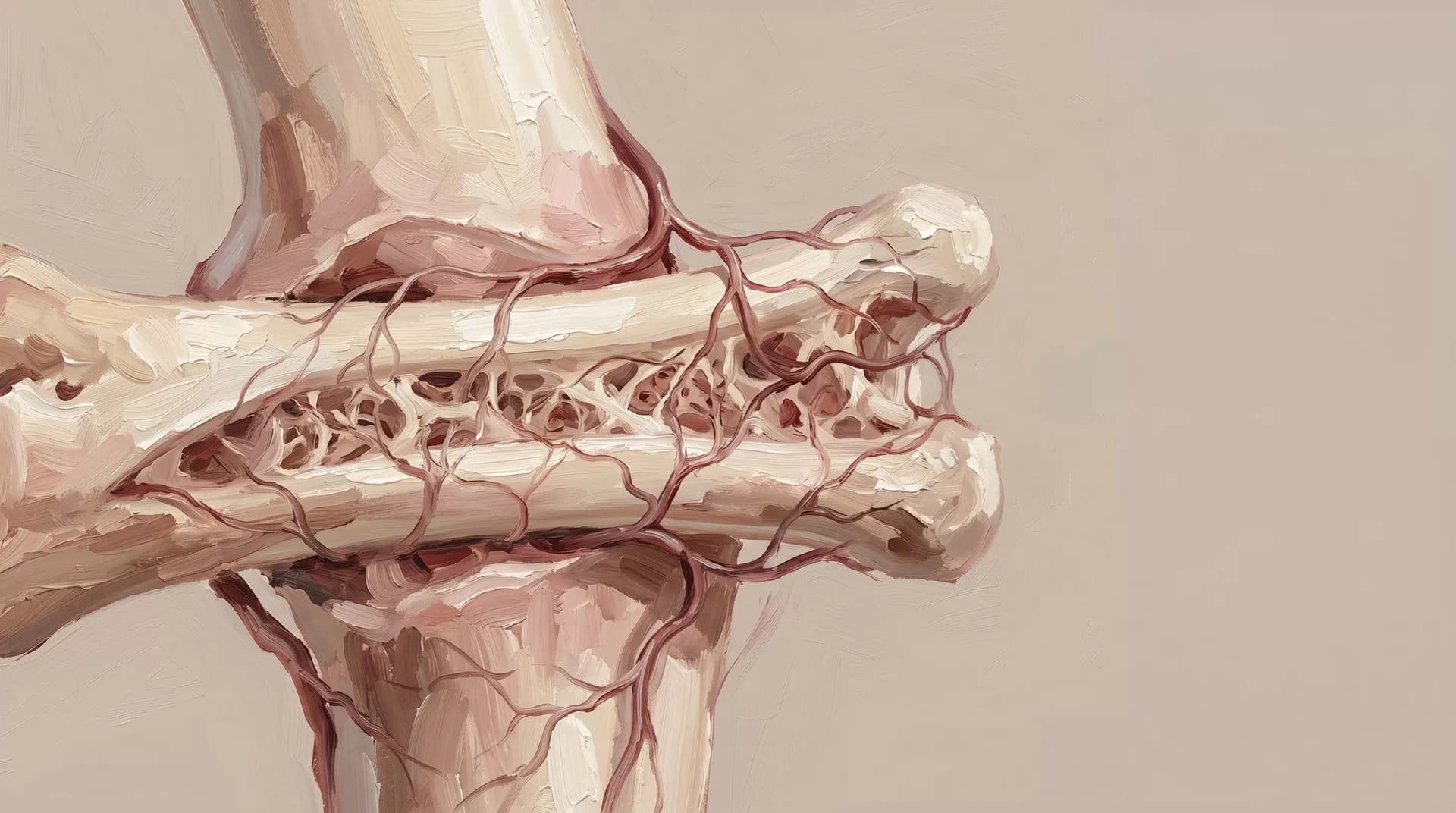
Revascularized bone grafting reconstructs maxillofacial defects using free bone transplants with microsurgical vascular anastomosis. ъ
Available sections:
Revascularized Bone Grafting
Revascularized bone grafting is a method of restoring bone defects in which a living bone graft with its own blood supply is transplanted to the area of lost or resected bone. Unlike conventional bone grafting, where a bone block without vessels is used, a revascularized graft is transferred together with its feeding artery and vein. After transfer, the vessels are sutured to the vessels of the recipient area under an operating microscope.
Continuous blood flow makes this bone viable: it does not resorb, integrates reliably, and preserves its volume in the long term. At Iskusstvo Clinic in Moscow, this method is used as part of comprehensive reconstruction of the maxillofacial region, including with subsequent dental implant placement.
This page is dedicated exclusively to bone reconstruction. If the defect involves only soft tissues, the description of soft tissue flaps is available on the Free Flap Transfer Using Microsurgical Technique page.
Indications
Defects of the lower jaw after resection due to tumors
Defects of the upper jaw after oncological surgery
Post-traumatic defects of the facial skeleton with significant bone volume loss
Osteomyelitis with sequestration and extensive bone destruction
Avascular necrosis of the facial bones, including after radiation therapy or bisphosphonate therapy
Reconstruction in an irradiated area where local tissues cannot ensure integration of a conventional graft
Defects with planned dental implant placement requiring the creation of a full bone foundation
Cases in which previous bone grafting methods have not provided a result
Donor Areas
The choice of donor site depends on the shape and volume of the defect, the need for bone modeling, and plans for further rehabilitation.
Fibula Free Flap
The fibula is the most commonly used source of bone material in maxillofacial surgery. It has sufficient length and cortical strength and can be modeled well. It can be used to recreate the curves of the lower or upper jaw.
The flap includes a soft tissue component, which can simultaneously close a soft tissue defect. Later, the fibula allows dental implants to be placed for prosthetic rehabilitation.
The scar is located on the lower leg. With proper technique, limb function is usually not significantly affected.
Radial Osteocutaneous Flap
This is a thinner bone fragment used for subtotal defects of the upper jaw and situations where a small bone volume is needed in combination with thin soft tissue coverage.
The donor area is the forearm.
Iliac Crest
The iliac crest provides a large volume of cancellous bone. It is used for reconstruction of large defects with a pronounced bone volume deficit.
How the Operation Is Performed
Preoperative Planning
CT imaging with 3D reconstruction of the defect area is performed, along with angiography or Doppler ultrasound of the donor area vessels.
In complex cases, individual stereolithographic templates are made to reproduce bone structures accurately. When dental implant placement is planned, the required volume and spatial position of the bone block are calculated in advance.
Anesthesia
The operation is performed under general anesthesia. The duration and nature of anesthesiology support are planned with the long procedure in mind.
Course of Surgery
Two surgical teams work simultaneously: one prepares the recipient area, including resection and identification of recipient vessels, usually the facial artery and vein or the superior thyroid artery; the other harvests the bone flap.
The bone block is shaped according to the defect using templates and fixed with titanium plates. After bone fixation, microvascular anastomosis is performed under the operating microscope.
Duration
The operation lasts from 5 to 12 hours, depending on the scope. Exact timing is determined during preoperative planning.
Result and Rehabilitation
The first 48–72 hours after surgery are the critical period for monitoring graft viability. Hospitalization usually lasts from 10 to 14 days.
Approximate recovery timelines:
Primary healing of soft tissues takes 3–4 weeks
Consolidation, or integration, of the bone graft takes 3 to 6 months
Dental implant placement, when planned, is usually performed 4–6 months after consolidation is confirmed
All timelines are individual and depend on the scope of reconstruction, tissue condition, and the course of healing. The donor area, such as the lower leg in a fibula flap, requires monitoring of foot function during the early postoperative period.
Contraindications
Contraindications include severe systemic diseases in the stage of decompensation, pronounced vascular atherosclerosis affecting the planned donor or recipient arteries, and uncorrectable blood clotting disorders.
Oncological disease is not an absolute contraindication. The decision is made jointly with the oncologist.
The final conclusion on operability is made by the surgeon after a complete examination.
Frequently Asked Questions
How Is Revascularized Bone Grafting Different from Conventional Bone Grafting?
In conventional bone grafting, a bone block without vessels is transplanted. It integrates through the ingrowth of vessels from the surrounding tissues. This works for small defects in well-vascularized areas.
In large defects, irradiated tissues, or scar-altered tissues, such bone may fail to integrate or may resorb. A revascularized graft brings its own blood flow and works where conventional grafting is not possible.
Can Dental Implants Be Placed After Reconstruction?
In most cases, yes. Revascularized bone creates a full bone foundation suitable for implant placement.
The timing and specific possibility depend on the scope of reconstruction and the quality of graft integration. These details are discussed individually.
Will the Leg Be Affected After Fibula Harvesting?
The fibula does not carry the main weight-bearing load. With proper technique, limb function is not significantly impaired in most patients.
During the early postoperative period, monitoring is performed and, if necessary, rehabilitation exercises are prescribed. A scar remains on the lower leg.
Is the Operation Always Performed in Several Stages?
Not always. In oncological diseases, tumor removal and reconstruction are often performed at the same time. This helps avoid a period with an open defect.
In other situations, reconstruction is planned as a delayed procedure. The treatment strategy is determined by the surgeon and, if necessary, together with the oncologist.
How Long Does Recovery Take?
The inpatient stage usually lasts 10–14 days. Return to everyday life usually takes 4–6 weeks, depending on the scope of surgery.
Full bone consolidation and the final result form over 6–12 months.
Book a Consultation
Book a consultation. The surgeon will assess the size of the defect, select the donor area, and discuss the stages of reconstruction.
OUR SPECIALISTS
A team that continues the traditions of Professor A.I. Nerobeev's school. Our specialists not only practice but also teach, developing unique techniques in reconstructive and aesthetic medicine.

Leave your number and we will contact you