 My Account
 +7 (495) 129 37 00
 st. Derbenevskaya, 9
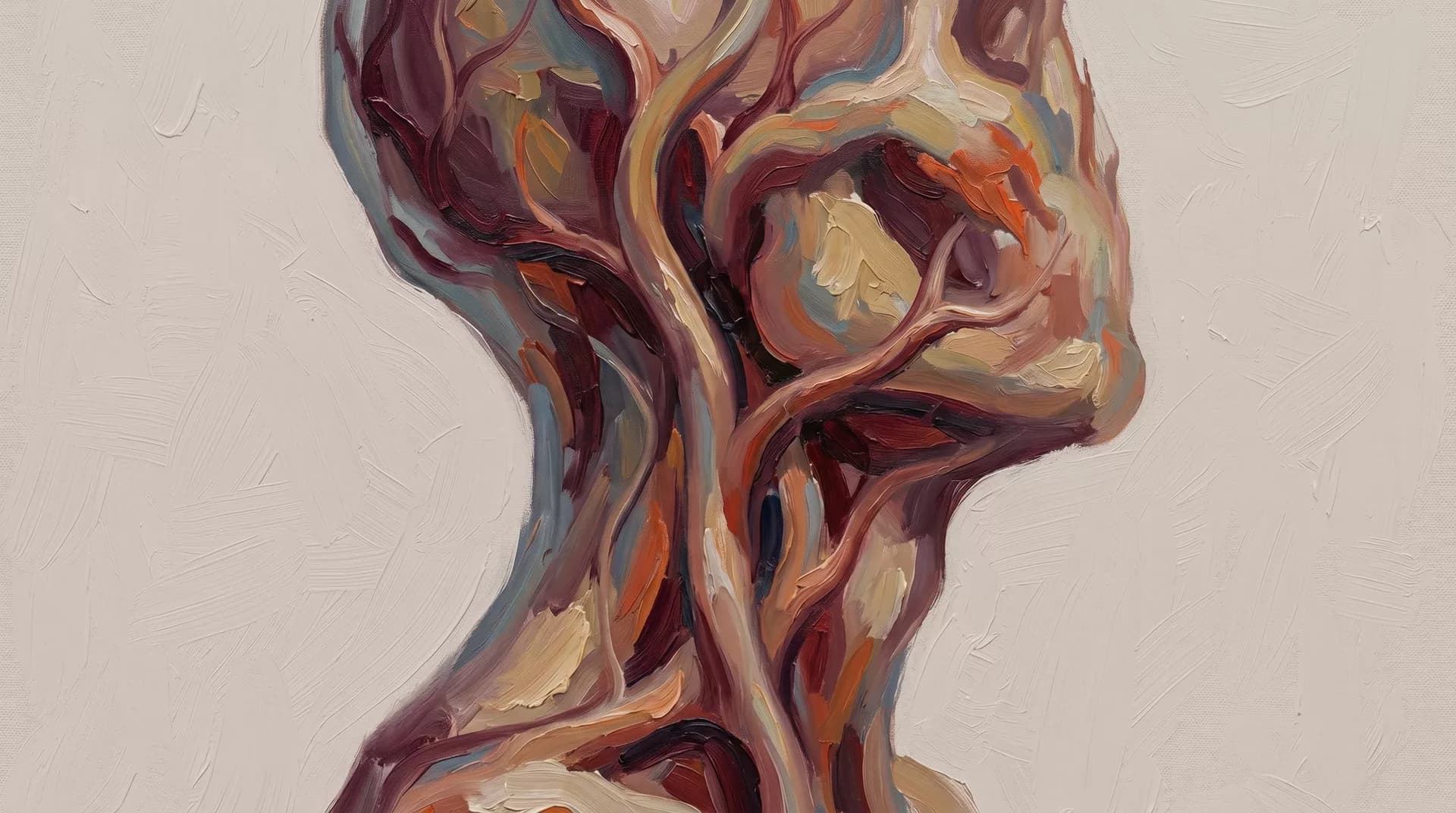
vascular-lesions-treatment
Removal of vascular lesions with laser and sclerotherapy.
Available sections:
Treatment of Vascular Lesions of the Head and Neck
Malformations, Telangiectasias, and Chemodectomas
Vascular lesions of the head and neck are a broad group of conditions that include congenital vascular anomalies, or malformations, and vascular tumors. They differ in origin, behavior, and treatment methods, which is why precise diagnostics are required before selecting a treatment strategy.
Some lesions grow slowly and may not cause discomfort for years. Others progress, deform tissues, cause bleeding, or impair function. At Iskusstvo Clinic in Moscow, treatment of vascular lesions is planned individually, taking into account the type, size, and location of the lesion.
Types of Vascular Lesions
Venous Malformations
Venous malformations are congenital anomalies of the venous vessels. They are soft, bluish lesions that compress when pressed. They do not regress on their own and gradually increase in size.
Treatment methods include sclerotherapy, meaning injection of a sclerosing agent into the malformation, and/or surgical excision. The choice depends on the size and location of the lesion.
Arteriovenous Malformations, or AVMs
This is the most complex type of vascular lesion: an abnormal connection between arteries and veins without a capillary bed. These lesions pulsate, feel warm to the touch, and are prone to growth and bleeding.
Treatment involves surgical excision, often with preliminary embolization, meaning occlusion of the feeding vessels, to reduce blood loss. The operation is technically complex and requires careful preoperative planning.
Lymphatic Malformations
These are cystic lesions arising from lymphatic vessels, most often in the neck and submandibular area. They may reach significant size and compress the airway.
Treatment includes sclerotherapy, which is especially effective for macrocystic forms, or surgical excision.
Telangiectasias
Telangiectasias are dilated superficial skin vessels, often described as “spider veins” or “vascular stars.” On the face, they are most often treated with laser methods in the cosmetology department. Extensive lesions may be treated within the framework of surgical care.
Chemodectomas, or Paragangliomas
Chemodectomas are rare benign tumors arising from chemoreceptor tissue. In the head and neck area, the most common types are carotid body tumor, located at the bifurcation of the carotid arteries, and jugular glomus tumor.
They grow slowly, but may reach large sizes and compress vessels and nerves. Treatment is surgical removal, often requiring the participation of a vascular surgeon.
Diagnostics
Accurate diagnosis of the type of vascular lesion is the mandatory first step. Ultrasound with Doppler imaging is used to assess blood flow. Contrast-enhanced MRI is used to determine the extent of the lesion. Angiography is used for arteriovenous malformations and chemodectomas.
Based on the examination results, the surgeon selects the method and scope of treatment.
Treatment Methods
Sclerotherapy
Sclerotherapy involves injecting a sclerosing agent directly into the cavity of the malformation under ultrasound or fluoroscopic guidance. This causes obliteration, or closure, of the abnormal vessels.
The method is most effective for venous and lymphatic malformations. Several sessions may be required.
Surgical Excision
Surgical excision is indicated for lesions that do not respond to sclerotherapy, in cases of recurrence, or when the function of surrounding structures is impaired.
In arteriovenous malformations, surgery is performed after embolization of the feeding vessels.
Combined Approach
For extensive lesions, the best result is achieved by combining sclerotherapy and surgery. Sclerotherapy reduces the size of the lesion and decreases intraoperative blood loss, while surgery removes the residual volume.
Rehabilitation
After sclerotherapy, outpatient follow-up is performed. Swelling in the treatment area resolves within 1–2 weeks.
After surgical removal, hospitalization lasts from 3 to 10 days, depending on the scope of surgery.
The final result forms within 3–6 months.
Contraindications
Contraindications depend on the treatment method. For sclerotherapy, they include allergy to the medication, blood clotting disorders, and pregnancy.
For surgery, general surgical contraindications apply.
The decision is made by the surgeon after a complete examination.
Frequently Asked Questions
How Is a Malformation Different from a Hemangioma?
A hemangioma is a vascular tumor that grows during the first months of life and then regresses on its own.
A malformation is a structural vascular defect that does not regress, but slowly progresses. The treatment strategies for these conditions are fundamentally different.
Is an Arteriovenous Malformation Dangerous?
An AVM is a potentially serious lesion. It tends to grow and may cause bleeding, including heavy bleeding.
Delaying treatment usually makes surgery more difficult. If an AVM is diagnosed, consultation with a surgeon is recommended without delay.
Is Surgery Needed If a Chemodectoma Is Small and Does Not Cause Discomfort?
Small carotid body tumors in young patients are recommended for removal while they are still small and do not surround the carotid arteries.
As the tumor grows, surgery becomes significantly more difficult. In elderly patients with small asymptomatic tumors, observation may be possible.
How Many Sclerotherapy Sessions Are Needed?
It depends on the size and type of malformation. On average, 2–4 sessions are required at intervals of 4–8 weeks.
The result is assessed using MRI or ultrasound. If the effect is insufficient, a surgical stage may be added.
Book a Consultation
Book a consultation. The surgeon will review the examination results and recommend the optimal treatment strategy.
OUR SPECIALISTS
A team that continues the traditions of Professor A.I. Nerobeev's school. Our specialists not only practice but also teach, developing unique techniques in reconstructive and aesthetic medicine.

Leave your number and we will contact you