 My Account
 +7 (495) 129 37 00
 st. Derbenevskaya, 9
Removal of Benign Formations
Laser, radio-surgical or cryodestructive removal of lipomas, atheromas, fibromas, papillomas and nevi on face, neck, body provides bloodless tissue vaporization without scars, bleeding or recurrence anywhere.
Skin Formation Removal (nevus, papilloma, kerathoma, dermatofibroma, angioma, etc.)
Skin Formation Removal (nevus, papilloma, kerathoma, dermatofibroma, angioma, etc.) Laser, radio wave or electrocoagulation removal of nevi, papillomas, keratomas, dermatofibromas and angiomas provides precise bloodless tissue vaporization without scars, bleeding or recurrence on face, neck and body.
Available sections:
Removal of benign lesions is a field of plastic surgery that includes surgical excision of skin and subcutaneous neoplasms: moles, or nevi, papillomas, keratomas, dermatofibromas, angiomas, lipomas, atheromas, and other lesions. At Iskusstvo Clinic in Moscow, lesion removal is performed by surgeons trained in the school of Professor Nerobeev, which ensures not only safe excision, but also an aesthetically optimal result, with minimal scarring planned according to the skin tension lines.
The key difference between surgical removal in a plastic surgery clinic and removal in a cosmetology office is mandatory histology, or morphological examination of the removed tissue, and wound closure techniques aimed at forming a minimally visible scar. Every removed lesion is sent for histological examination to rule out malignancy.
Indications
Medical Indications
Lesions suspicious for malignancy, including changes in color, shape, or size, bleeding, or rapid growth; surgical excision with histology is indicated
Lesions exposed to chronic trauma, such as those located along clothing lines, bra straps, belts, in skin folds, or on the scalp
Inflamed or infected lesions, including suppurated atheromas or inflamed nevi
Rapidly growing lesions requiring morphological verification
Aesthetic Indications
Lesions on exposed areas of the body, such as the face, neck, décolleté, and hands, causing aesthetic discomfort
Multiple papillomas, keratomas, or nevi that reduce quality of life
Large or protruding lesions that interfere with everyday life
Types of Lesions
Skin Lesions
Skin lesions are located within the thickness of the skin or on its surface. They include nevi, or moles, which are pigmented lesions formed by melanocytes; papillomas, which are soft stalk-like skin growths caused by HPV; keratomas, which are keratinizing lesions associated with age-related skin changes; dermatofibromas, which are dense nodules formed from connective tissue; and angiomas, which are red or burgundy vascular lesions.
Subcutaneous Lesions
Subcutaneous lesions are located in the subcutaneous fat tissue or deeper. They include lipomas, which are benign tumors of fatty tissue that are soft and mobile; atheromas, or epidermal cysts, which are sebaceous gland cysts with characteristic contents; and fibromas, which are connective tissue lesions.
Removal Methods
Surgical Excision
Surgical excision is the main method used at Iskusstvo Clinic. The lesion is removed with a scalpel as a single block with a margin of healthy tissue around the perimeter, usually 1–3 mm. This ensures complete removal and allows full histological examination.
The wound is closed layer by layer with a fine cosmetic suture, taking into account the skin tension lines, also known as Langer’s lines. This helps form a minimally visible scar. For facial lesions, especially fine sutures, such as 6-0 and 7-0, and microsurgical instruments are used.
Electrocoagulation and Radiofrequency Removal
These methods are used for small superficial lesions, such as papillomas, small keratomas, and flat nevi. The lesion is removed using electric current or radiofrequency energy. The method does not require sutures, but it does not provide sufficient material for histology in cases of deeper lesions.
Laser Removal
Laser removal is used for superficial vascular and pigmented lesions. Its advantage is minimal trauma to the surrounding tissues. The limitation is that it does not always allow tissue to be obtained for histological examination.
How the Procedure Is Performed
Before removal, the surgeon examines the lesion and, if necessary, performs dermoscopy, which is examination under magnification using a dermatoscope. The optimal removal method and incision direction are then determined.
The procedure is performed under local anesthesia. Duration ranges from 10 to 30 minutes for one lesion; for multiple lesions, it may take up to 1–1.5 hours. The patient leaves the clinic on the same day.
The removed tissue is labeled and sent for histological examination to a pathomorphology laboratory. Results are usually ready within 7–14 days. If histology reveals malignant changes, the surgeon, together with an oncologist, determines the further treatment plan, which may include additional excision or observation.
Rehabilitation
Sutures are removed on days 5–7 for the face and days 10–14 for the body. The wound is treated with an antiseptic daily. Physical activity involving the wound area is restricted for 2–3 weeks.
Sunscreen must be applied to the scar for 6–12 months to prevent hyperpigmentation. The scar matures over 3–6 months on the face and 6–12 months on the body, gradually becoming lighter.
Risks and Complications
Possible complications include bleeding, infection, hypertrophic or keloid scarring, hypo- or hyperpigmentation, and recurrence of the lesion in cases of incomplete removal.
With surgical excision and complete removal, recurrence is extremely rare. The risk of keloid scarring depends on individual predisposition and location. The sternum and earlobe areas are considered higher-risk zones.
Contraindications
Acute infectious diseases
Blood clotting disorders
Active inflammation in the planned removal area; this is a relative contraindication and may require preliminary treatment
Pregnancy; this is a relative contraindication, planned removal is usually postponed, while urgent removal may be performed
The final decision is made by the doctor.
Why Choose Iskusstvo Clinic
Plastic Surgery Technique
Excision and wound closure are performed according to the principles of plastic surgery, including Langer’s lines, layered closure, and fine suture material. This helps achieve minimally visible scars.
Mandatory Histology
Every removed lesion is examined morphologically to rule out malignant changes.
Microsurgical Expertise
For facial lesions, microsurgical instruments and optical magnification are used.
Comprehensive Approach
If necessary, lesion removal can be combined with other procedures, such as blepharoplasty or scar correction.
Frequently Asked Questions
Is It Always Necessary to Remove a Benign Lesion?
Not all lesions require removal. Indications include suspicion of malignancy, chronic trauma, rapid growth, and aesthetic discomfort. Stable lesions that do not cause concern may be monitored.
The decision is made individually during the consultation.
Why Is Histology Necessary?
Histological examination is the only reliable way to confirm that a lesion is benign. Visual assessment, even with dermoscopy, does not provide a 100% guarantee.
Histology helps rule out skin cancer, including basal cell carcinoma, squamous cell carcinoma, and melanoma, which is critically important for patient safety.
Will There Be a Scar After Removal?
A scar remains after any surgical procedure, but its visibility depends on the closure technique, incision placement, and individual characteristics.
The surgeons at Iskusstvo Clinic place the incision along the skin tension lines and close the wound layer by layer, which helps form a thin, minimally visible scar. Facial scars mature faster and usually become almost invisible within 3–6 months.
Can Several Lesions Be Removed in One Visit?
Yes, provided that the total scope of the procedure allows it. In one visit, it is possible to remove 5–15 small lesions, such as papillomas or keratomas, or 2–5 larger lesions, such as nevi, atheromas, or lipomas.
The exact number is determined by the surgeon, taking into account the location, size, and patient comfort.
When Is Urgent Medical Attention Needed?
Urgent consultation is required if the lesion has increased rapidly over the past few weeks, changed color, become darker or unevenly colored, started bleeding, developed an ulcer on the surface, become painful despite previously being painless, or if new lesions appear around an existing one.
These signs require examination by a surgeon and, possibly, urgent biopsy.
Book a consultation. The surgeon will examine the lesion, perform dermoscopy if necessary, and recommend the optimal removal method with mandatory histological examination.
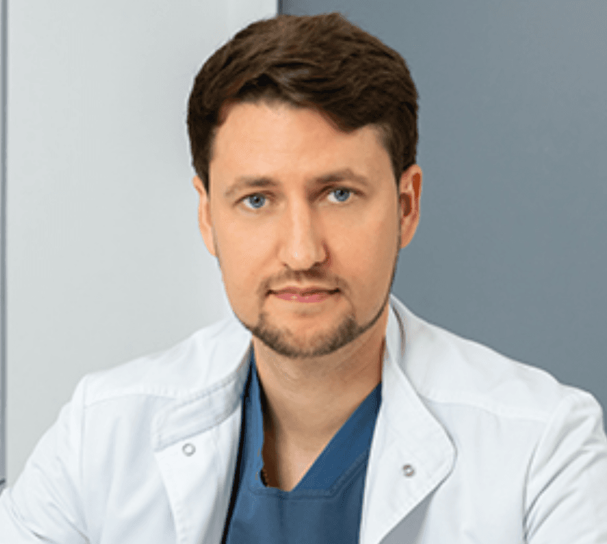
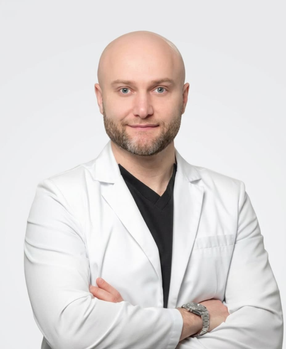
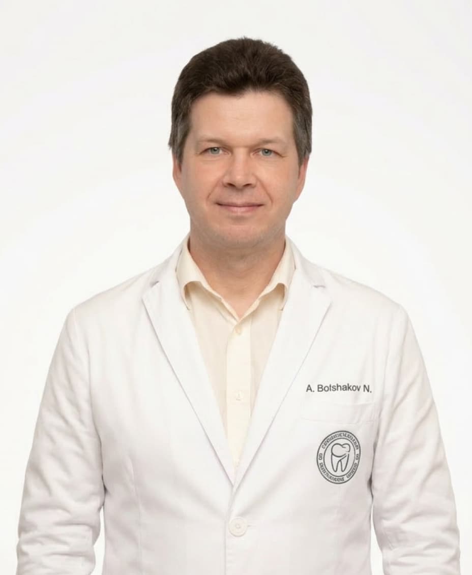
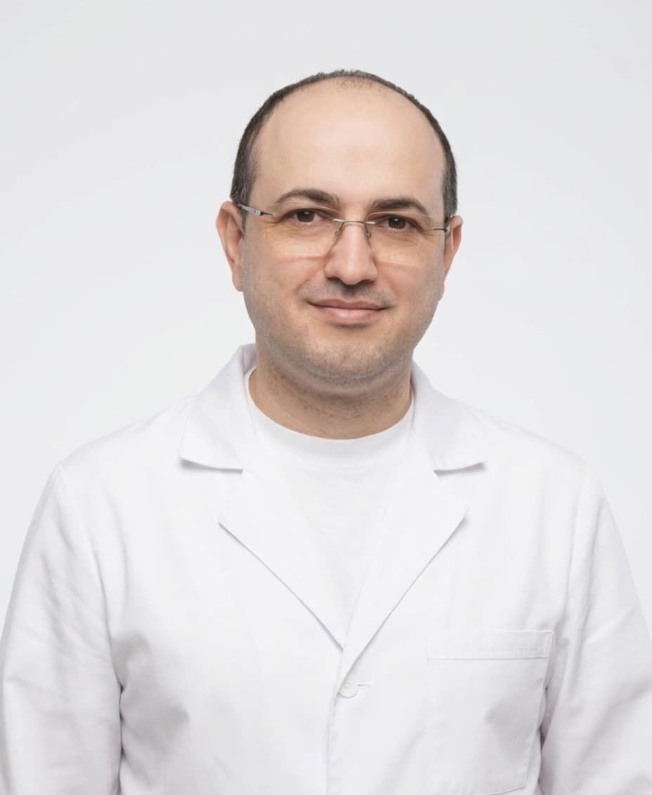
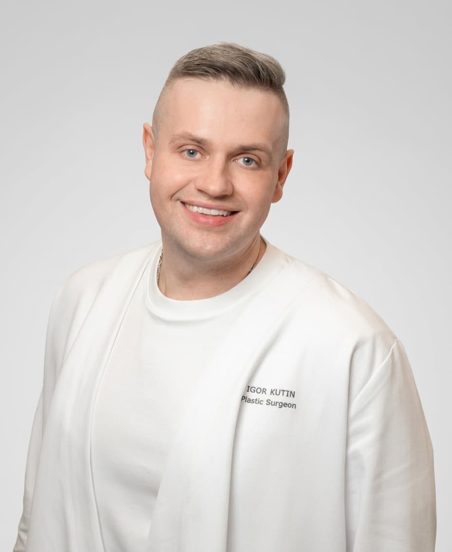
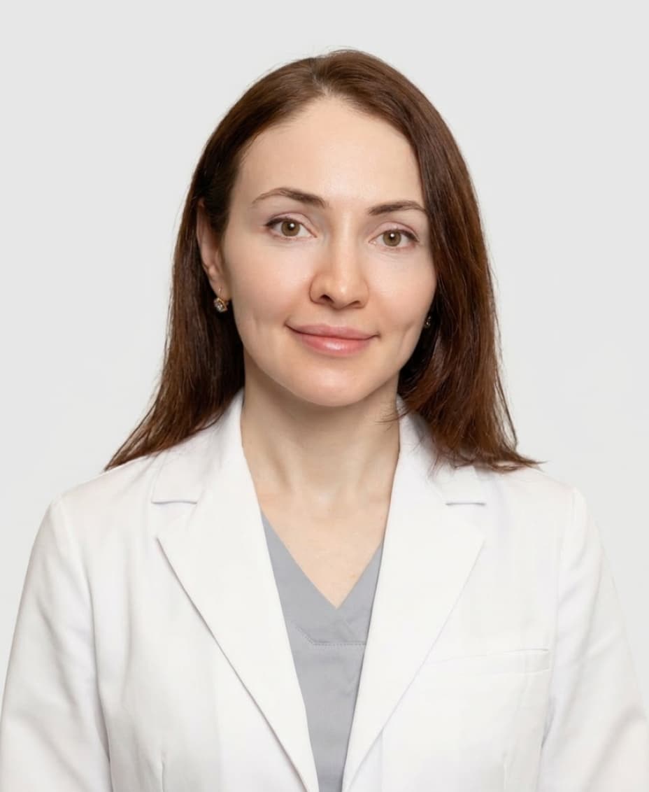
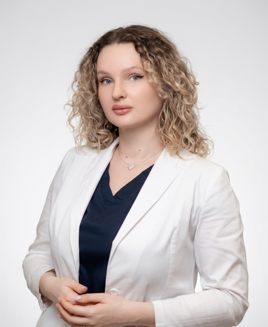
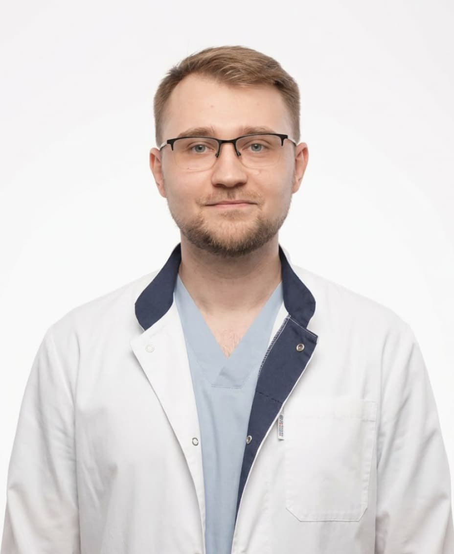
OUR SPECIALISTS
A team that continues the traditions of Professor A.I. Nerobeev's school. Our specialists not only practice but also teach, developing unique techniques in reconstructive and aesthetic medicine.


Leave your number and we will contact you