 My Account
 +7 (495) 129 37 00
 st. Derbenevskaya, 9
Tooth Diagnosis
Oral сavity examinations.
Available sections:
Dental diagnostics is the first stage of dental treatment. It helps the dentist identify not only visible problems but also hidden changes affecting the teeth, roots, bone tissue, bite, and temporomandibular joints (TMJ).
Even if a patient is concerned about a single tooth, the source of pain or discomfort may be deeper: decay beneath an old filling, inflammation around the root, a crack, excessive loading, a bite disorder, a periodontal pocket, or a problem involving a neighboring tooth. Without proper diagnostics, it is difficult to create an accurate and safe treatment plan.
At Iskusstvo Clinic, dental diagnostics is performed before restorative treatment, implant placement, prosthetic treatment, orthodontics, surgical procedures, and cosmetic dentistry. The dentist selects the most appropriate diagnostic method depending on the clinical situation: a preventive examination, periapical X-ray, panoramic X-ray, CBCT scan, intraoral 3D scanning, smile design, or gnathological assessment.
The goal of diagnostics is not simply to “find a problem,” but to understand its cause, evaluate the prognosis of the teeth, and select treatment that is justified, precise, and safe.
When Dental Diagnostics Is Recommended
Dental diagnostics may be recommended in the following situations:
• Tooth pain or sensitivity.
• Suspected dental caries, pulpitis, or periodontitis.
• Before dental implant placement.
• Before treatment with crowns, veneers, inlays, or bridges.
• Before orthodontic treatment.
• Before tooth extraction, especially for impacted or malpositioned teeth.
• In cases of gum inflammation or periodontal pockets.
• Tooth wear and bruxism.
• Clicking, pain, or discomfort in the TMJ.
• Preventive monitoring of the condition of the teeth and gums.
Diagnostic Methods
Preventive Dental Examination
A preventive examination helps identify cavities, gum inflammation, aging restorations, chips, cracks, tooth wear, and other changes at an early stage.
The dentist evaluates the condition of the teeth, oral mucosa, gums, bite, and existing fillings, crowns, or implants. Additional diagnostic procedures may be recommended if necessary.
Periapical Dental X-ray
A periapical X-ray is used when detailed evaluation of one or several teeth is required. It helps assess the condition of the root, root canals, tissues surrounding the root apex, the depth of decay, and the quality of previous treatment.
This type of imaging is commonly prescribed for pain, suspected inflammation, before root canal treatment, or after endodontic therapy.
Panoramic Dental X-ray
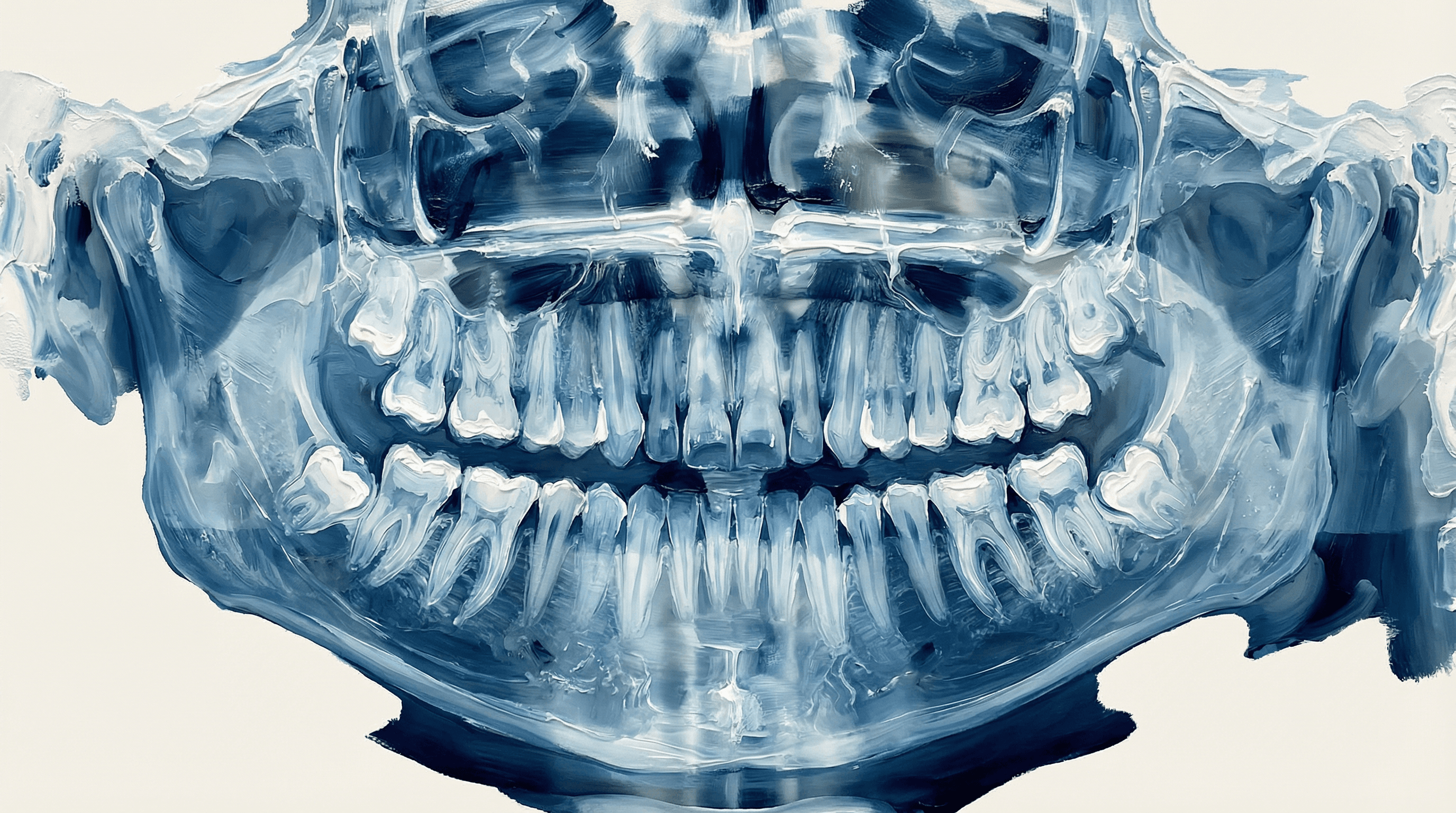
A panoramic X-ray provides a comprehensive image of the entire dentofacial system, including teeth, roots, developing teeth, jaws, maxillary sinuses, and the temporomandibular joint region.
It is commonly used as an overview examination before comprehensive treatment, orthodontics, tooth extraction, implant placement, and assessment of overall oral health.
CBCT Scan
Cone Beam Computed Tomography (CBCT) is a three-dimensional imaging study of the teeth and jaws. It allows the dentist to evaluate bone volume, root position, canal anatomy, inflammatory lesions, impacted teeth, and proximity to important anatomical structures.
CBCT is particularly important before implant placement, complex extractions, bone grafting procedures, apical surgery, and treatment of complex root canal systems.
Intraoral 3D Scanning
Intraoral 3D scanning creates a digital model of the teeth without the need for traditional impressions.
It is used for prosthetic treatment, crowns, veneers, inlays, aligners, night guards, retainers, and diagnostic planning. Digital scanning allows the dentist and dental laboratory to work with greater accuracy and comfort for the patient.
Smile Design
Smile design is used in cosmetic dentistry to plan the future appearance of a patient’s smile in advance.
The dentist can evaluate the shape, length, proportions, and overall aesthetics of the future smile. This method is especially useful before veneer treatment, cosmetic restorations, prosthetic rehabilitation in the smile zone, and comprehensive smile makeovers.
Gnathological Assessment
Gnathological diagnostics evaluates not only the teeth but also the function of the entire masticatory system, including the bite, mandibular movements, chewing muscles, and temporomandibular joints.
It is recommended for patients with bruxism, tooth wear, TMJ pain, clicking sounds, tension headaches, and before complex prosthetic treatment or changes in bite height.
How Diagnostics Is Performed
The process begins with a discussion of the patient's complaints and medical history. The dentist asks about symptoms, when they began, and any previous treatment, trauma, tooth extractions, orthodontic treatment, implants, or prosthetic work.
Next, a clinical examination of the oral cavity is performed. The dentist evaluates the teeth, gums, oral mucosa, bite, restorations, crowns, implants, and oral hygiene.
Additional diagnostic procedures are then selected as needed. In some cases, a clinical examination and periapical X-ray are sufficient. In others, CBCT imaging, digital scanning, photographic documentation, or gnathological assessment may be required.
Based on the diagnostic findings, the dentist explains the situation to the patient, identifies problem areas, and develops an individualized treatment plan.
Why Comprehensive Diagnostics Matters
Comprehensive diagnostics helps avoid treatment based on assumptions.
For example, before placing a crown, it is important to evaluate not only the shape of the tooth but also the condition of the root, root canals, gums, and bite. Before implant placement, bone volume and the location of important anatomical structures must be assessed. Before veneer treatment, the dentist should evaluate the bite, enamel quality, facial proportions, and smile line.
The more accurate the diagnostics, the more reliable and predictable the treatment plan becomes, and the lower the risk of unexpected complications during subsequent stages of care.
OUR SPECIALISTS
A team that continues the traditions of Professor A.I. Nerobeev's school. Our specialists not only practice but also teach, developing unique techniques in reconstructive and aesthetic medicine.




Leave your number and we will contact you